|
|
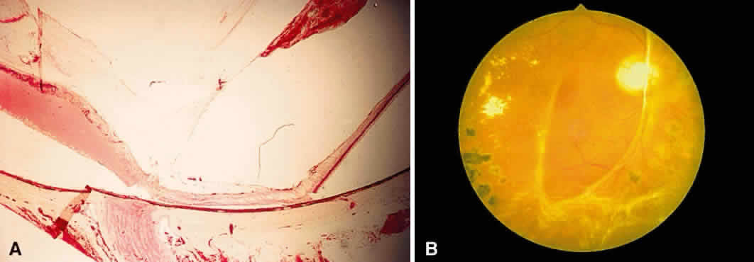
| Fig. 17. A. A histologic section stained with H & E demonstrating vascularized membranes on the disc and retina. Tractional retinal detachment is present. Notice the subretinal fluid (amorphous eosinophilic material). B. Fundus photograph of fibrous tissue emanating from the disc in patient with proliferative diabetic retinopathy. The disc is pale; the macula is edematous with exudate. Pigmented laser spots are visible temporally. |