|
|
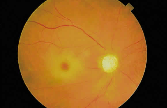
| Fig. 8. Fundus photograph of “cherry-red spot” in a patient with central retinal artery occlusion. The perifoveal edema (white opacification) does not extend beyond the macula because of the reduced concentration of ganglion cells in the extramacular retina. Marked pallor of the optic disc is present. |