|
|
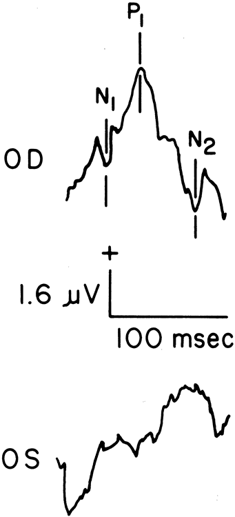
| Fig. 8. Pattern electroretinograms (PERGs) elicited by 4 Hz (eight reversals per second) from a 37-year-old white female patient with pseudotumor cerebri of the left eye. The upper tracing is a normal PERG from the right eye. Note that N2 (N95) is larger than P1 (P50). The PERG in the lower tracing, from the affected left eye, is severely reduced. PERGs were recorded using 64 samples and an outer canthal reference. |