|
|
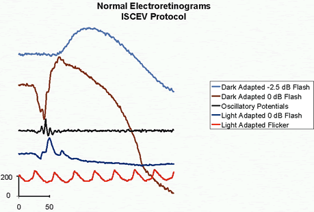
| Fig. 6. Electroretinograms (ERG) of a normal subject recorded using the ISCEV protocol. A. At the top, the light blue curve is a dark-adapted ERG elicited by a white light of 1.72 cd·m-2·s-1 attenuated by a 2.5 log unit neutral density filter. The subject had been dark adapted for 20 minutes. B. The second, brown waveform is a dark-adapted ERG elicited by a white light of 1.72 cd·m-2·s-1. C. The third, black wave shows dark-adapted ERG oscillatory potentials elicited by a white light with a luminance of 1.72 cd·m-2·s-1. D. The fourth, dark-blue waveform is a light-adapted ERG elicited by l-Hz flashes of 1.72 cd·m-2·s-1 against a 22 cd·m-2 background. E. ERGs elicited by 30-Hz flickering white light of 1.72 cd·m-2·s-1 against a 22 cd·m-2 background. |