|
|
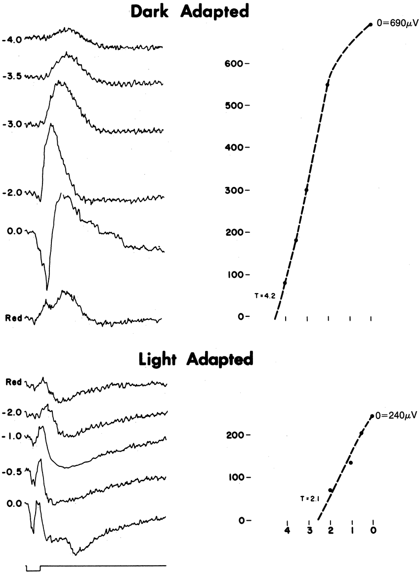
| Fig. 5. Electroretinogram (ERG) responses of a normal subject. We present a series of ERGs elicited by flashes of increasing intensity in the light and in the dark. On the left are responses obtained from the dark-adapted eye with increasingly brighter stimuli, measured in log units. The response to red stimuli shows a double-humped b wave in the light-adapted eye. An intensity response curve is shown on the right side of the diagram. The threshold value (T) shows where each curve crosses the arbitrarily chosen 50 μV level. One might also fit such functions with a Naka-Rushton or saturation function. |