|
|
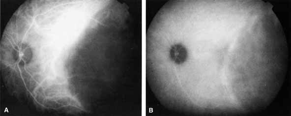
| Fig. 14 ICGA of a dome-shaped choroidal metastasis located in the temporal macular area. Both early phase (A) and middle-phase (B) photographs show relative hypofluorescence without angiographically apparent lesional vascularity. (Courtesy of Dr. J. Shields) |