|
|
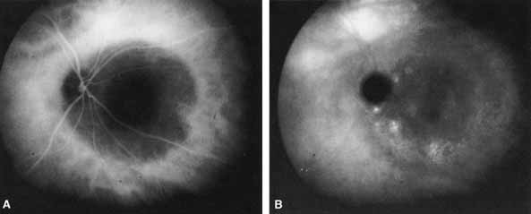
| Fig. 12 A deeply pigmented, dome-shaped choroidal melanoma in the nasal peripapillary area of the right eye. Early phase (A) and late-phase (B) ICG angiograms show the lesion to be relatively hypofluorescent with only punctate hyperfluorescent foci evident at the margins in the late phase. (Courtesy of Dr. J. Shields) |