|
|
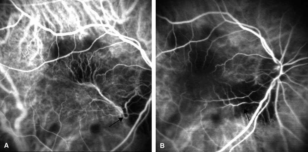
| Fig. 11 A. Pretreatment scanning laser ophthalmoscope high-speed indocyanine green angiography showing a well-delineated choroidal “feeder” (arrow) vessel supplying a large subfoveal neovascular complex. B. Posttreatment SLO-ICGA with laser photocoagulation applied to the feeder only (arrow) shows a lack of blood flow through the entire neovascular complex. |