|
|
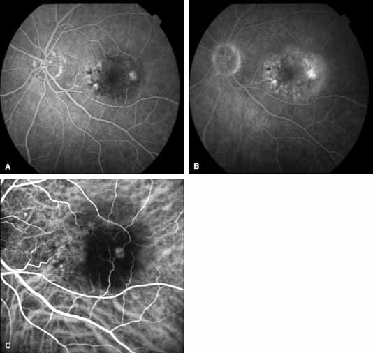
| Fig. 10 A. Early phase fluorescein angiogram shows a focal area of hyperfluorescence in the temporal macula. B. Late-phase fluorescein angiogram demonstrates diffuse, stippled hyperfluorescence consistent with occult neovascularization. C. SLO-based ICG demonstrates a “hot spot” with retinal vascular communication consistent with RAP. |