|
|
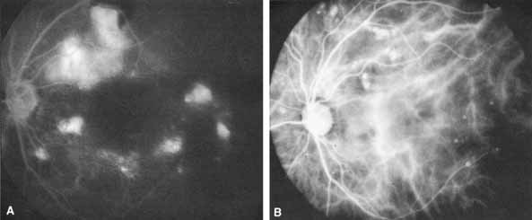
| Fig. 9 A case of idiopathic polypoidal choroidal vasculopathy with active subretinal exudation and hemorrhage superotemporal to the optic nerve head. A. A relatively late-phase fluorescein angiogram photograph of the involved left eye shows multiple areas of ill-defined subretinal fluorescein leakage and some blocked fluorescence along the superotemporal arcade from subretinal hemorrhage. B. Early phase ICG angiogram reveals multiple, bright hyperfluorescent foci clustered along choroidal vessels. These foci are concentrated superotemporal to the optic nerve in the area of clinically apparent exudation. |