|
|
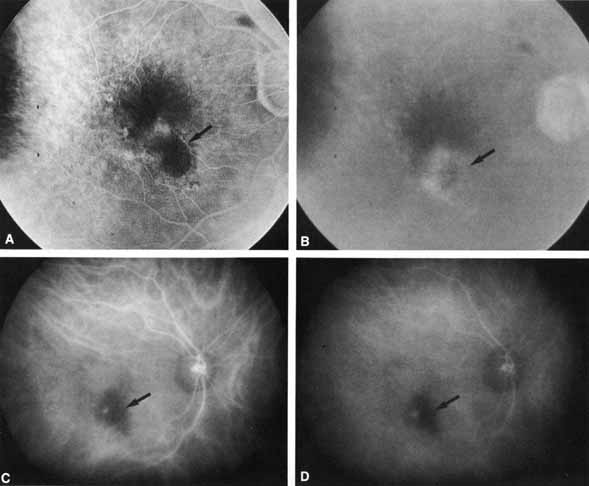
| Fig. 8 Early phase (A) and late-phase (B) fluorescein angiograms taken 2 weeks after laser photocoagulation of a choroidal neovascular focus in a patient with macular degeneration. There is ill-defined leakage (hyperfluorescence) along the temporal and superior aspect of the photocoagulation mark (arrow). Both early phase (C) and middle-phase (D) ICG angiograms demonstrate a small, well-defined focus of relative hyperfluorescence standing out in contrast at the margin of the hypofluorescent photocoagulation mark (arrow). Retreatment with laser photocoagulation directed at this hyperfluorescent spot alone resulted in rapid resolution of exudation. |