|
|
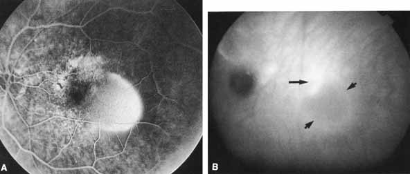
| Fig. 7 A. Arteriovenous phase fluorescein angiogram shows a bright, homogeneous hyperfluorescent area typical of a serous pigment epithelial detachment located temporal to the foveal center. The truncated nasal border of the detachment along with the adjacent mottled hyperfluorescence is suggestive of associated occult choroidal neovascularization in this patient with macular degeneration. B. The corresponding late-phase (20-minute) ICG angiogram reveals a bright hyperfluorescent area superotemporal to fixation (arrow) presumably representing the underlying choroidal neovascularization. The adjacent hypofluorescent serous pigment epithelial detachment (arrow heads) is seen in contrast. (Courtesy of Dr. E. Shakin) |