|
|
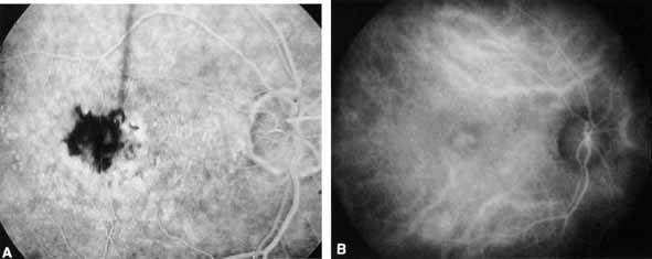
| Fig. 6 A. Arteriovenous phase fluorescein angiogram reveals a hypofluorescent area just temporal to fixation representing blockage from subretinal hemorrhage. Ill-defined hyperfluorescence is also seen along the nasal and inferior border of the blocked fluorescence, suggesting occult choroidal neovascularization in a patient with macular degeneration. B. A middle-phase (10-minute) ICG angiogram of the same eye demonstrates a discrete abnormal focus of relative hyperfluorescence corresponding to the area of subretinal hemorrhage. In contrast to the fluorescein angiogram, neither the thin layer of hemorrhage nor the fine drusen is evident on the ICG angiogram. |