|
|
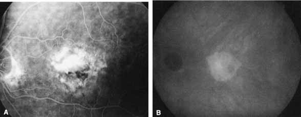
| Fig. 4 A. Arteriovenous phase fluorescein angiogram taken from a patient with macular degeneration shows leakage in the central macula along with surrounding ill-defined, speckled hyperfluorescence. B. The corresponding late-phase (25-minute) ICG angiogram demonstrates a well-defined, plaque-like area of relative hyperfluorescence. (Courtesy of Dr. W. Annesley) |