|
|
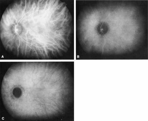
| Fig. 1 Normal Indocyanine Green (ICG) angiogram. A. Early phase photograph (90 seconds after dye injection) shows the hyperfluorescent choroidal and retinal vessels. B. Middle-phase photograph (10 minutes after dye injection) reveals more homogeneous background choroidal fluorescence with relative attenuation of the retinal vascular fluorescence. C. Late-phase photograph (30 minutes after dye injection) shows medium-sized choroidal vessels in relief (relative hypofluorescence). No retinal structures are visible. |