|
|
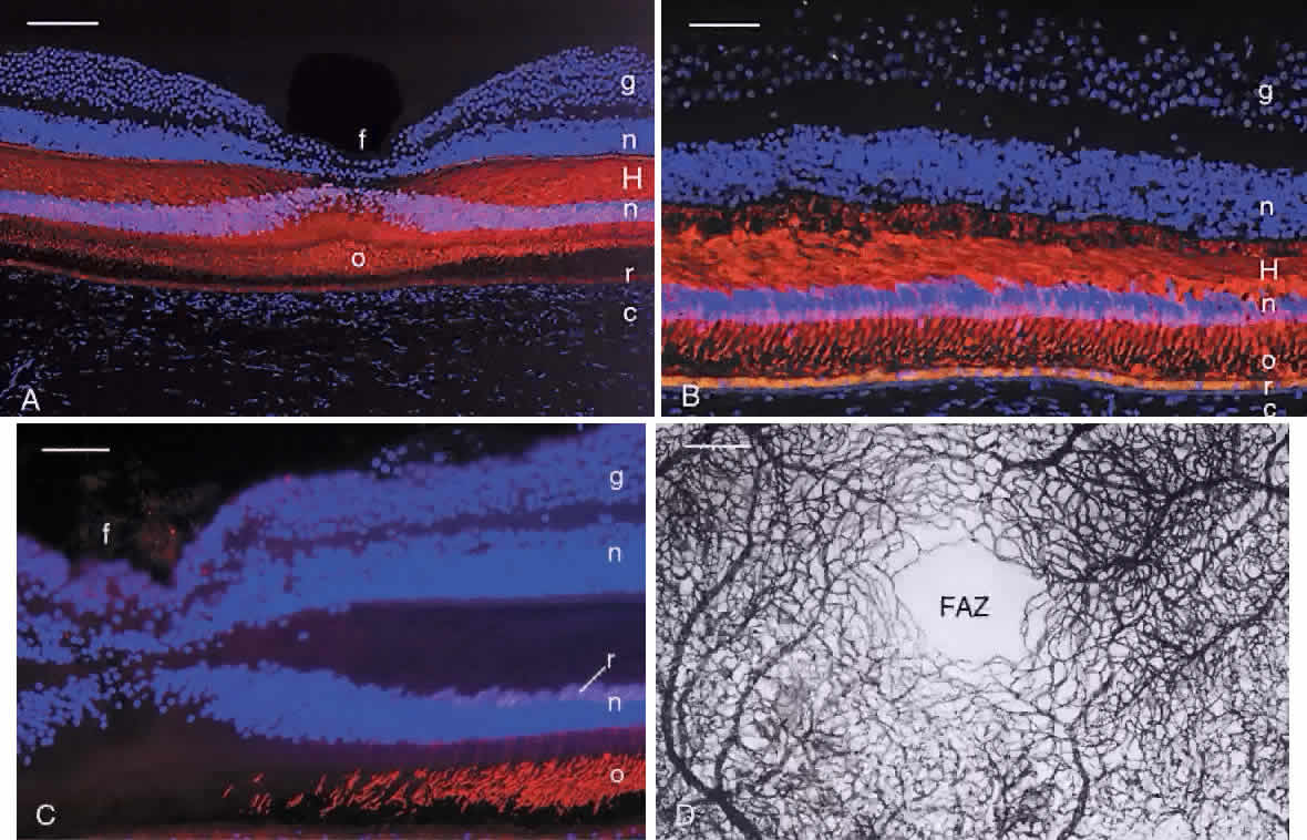
| Fig. 23. A. Immunocytochemical labeling of cones in human fovea (f) with monoclonal antibody 7G6 (red). Note tightly packed cones with long, thin outer segments (o). Cell nuclei (n) are counterstained blue with DAPI. c, choroid; r, retinal pigment epithelium; H, Henle fiber layer. Bar = 200 μm. B. Higher magnification of foveal cones labeled (red) with antibody 7G6. Note thick layer of photoreceptor axons that form the fiber layer of Henle (H). Cell nuclei (n) are counterstained blue with DAPI. The cone nuclei appear magenta because they are positive for 7G6. The rod nuclei internal to the cone nuclei in the outer nuclear layer are blue because they do not contain 7G6. o, outer segments; r, retinal pigment epithelium; c, choroid; g, ganglion cell layer. Bar = 100 μm. C. Immunocytochemical demonstration of scattered rods (rod outer segments [o] labeled red with antirhodopsin) in the wall of the fovea (f). The foveal cones are unlabeled. Cell nuclei (n) are counterstained blue with DAPI. g, ganglion cells; r, rhodopsin-positive rod cell bodies lying innermost in the outer nuclear layer. Bar = 50 μm. D. Capillary bed of the macula. The capillary-free zone (foveal avascular zone, FAZ) in the center is approximately 0.4 mm in diameter. Bar = 200 μm. (Modified from Hogan MJ, Alvarado JA, Weddell JE: Histology of the Human Eye. Philadelphia: WB Saunders, 1971.) |