|
|
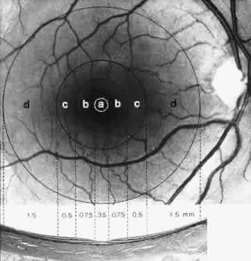
| Fig. 20. A fundus photograph is matched with a meridional light micrograph of the macular region. The fundus photograph shows the foveola (a), fovea (b), parafovea (c), and perifovea (d). (Hogan MJ, Alvarado JA, Weddell JE: Histology of the Human Eye. Philadelphia: WB Saunders, 1971.) |