|
|
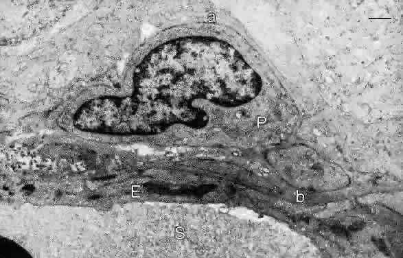
| Fig. 19. Electron micrograph of retinal capillary wall demonstrating an endothelial cell (E) and its basal lamina (b). A pericyte (P) is surrounded by a basal lamina (a) on the side facing the retinal parenchyma. S, serum in capillary lumen. Bar = 1.0 μm. (Hogan MJ, Alvarado JA, Weddell JE: Histology of the Human Eye. Philadelphia: WB Saunders, 1971.) |