|
|
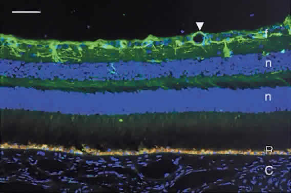
| Fig. 17. Immunofluorescence image of human retina demonstrating astrocytes labeled with antiglial fibrillary acidic protein (green). The astrocytes have long processes and are located mainly in the nerve fiber layer (f). The astrocytic foot processes surround the walls of a retinal blood vessel (arrowhead). R, autofluorescent lipofuscin granules in the retinal pigment epithelium; n, cell nuclei in outer and inner nuclear layers counterstained blue with DAPI. Bar = 50 μm. |