
|
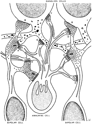
| Fig. 15. Schematic drawing of the synaptic interactions in the inner plexiform layer. A, B, and C depict bipolar synapses. D, E, and F represent amacrine cell contacts with bipolar and ganglion cells. Note ribbon-containing synapses formed by the bipolar cells. (Hogan MJ, Alvarado JA, Weddell JE: Histology of the Human Eye. Philadelphia: WB Saunders, 1971.) |