|
|
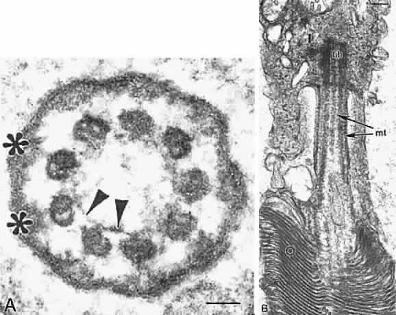
| Fig. 11. Electron microscopy of connecting cilia of human photoreceptors. A. Cross section of cilium. Note nine doublets of microtubules forming a ring characteristic of sensory cilia. The microtubules are interconnected by nexin links (arrowheads) and joined to the surface membrane by Y-shaped linkers (*). Bar = 0.04 μm. B. Longitudinal section of cilium. Note microtubules (mt), stacks of membrane discs in the outer segment (O), and basal body (bb) in the inner segment (I). Bar = 0.15 μm. |