|
|
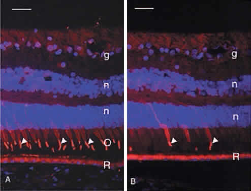
| Fig. 7. A. Immunocytochemical demonstration of red/green cone opsin (red, arrowheads) in the majority of human cone outer segments (O). R, retinal pigment epithelium; g, ganglion cells; n, DAPI-stained nuclei in the outer and inner nuclear layers. Bar = 30 μm. B. Immunocytochemical demonstration of blue cone opsin (red, arrowheads) in a minority of human cones. Arrowheads indicate the blue cone outer segments. R, retinal pigment epithelium; g, ganglion cells; n, DAPI-stained nuclei in the outer and inner nuclear layers. Bar = 30 μm. |