|
|
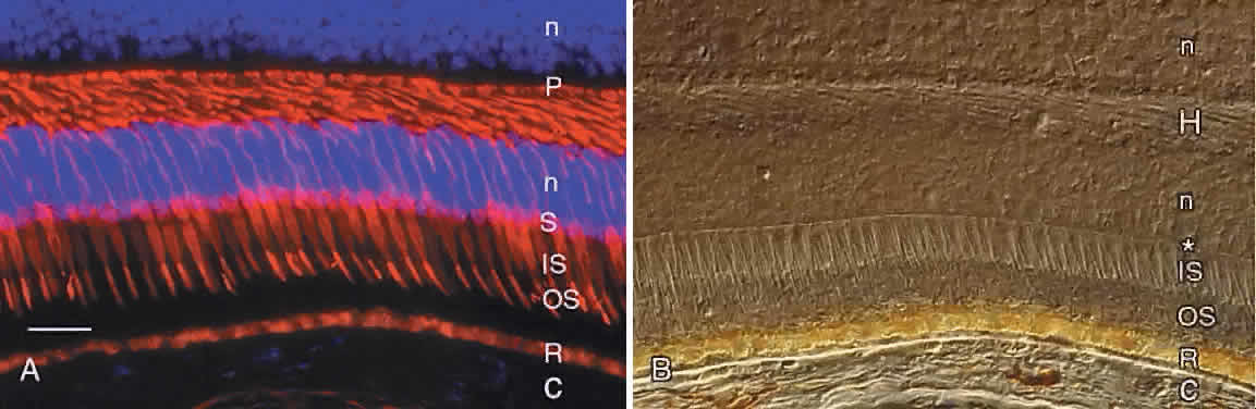
| Fig. 5. A. Immunocytochemical demonstration of human cone cytology by use of monoclonal antibody 7G6. Note the cone outer segments (OS), inner segments (IS), cell bodies (S), and synaptic pedicles (P). R, autofluorescent lipofuscin granules in the retinal pigment epithelium; C, choroid. Cell nuclei (n) are counterstained blue with DAPI. Bar = 30 μm. B. The same field as in A viewed by Nomarski differential interference contrast microscopy. Note external limiting membrane (*), photoreceptor outer segments (OS), and inner segments (IS). C, choroid; R, retinal pigment epithelium containing yellow lipofuscin and brown melanin granules; H, Henle fiber layer; n, nuclei in outer and inner nuclear layers. |