|
|
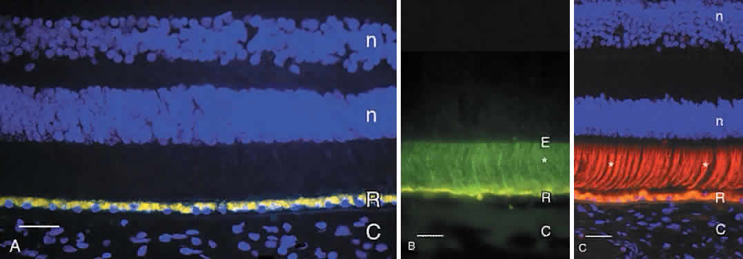
| Fig. 4. A. Fluorescence micrograph of adult human retina demonstrating autofluorescent lipofuscin granules that fill the retinal pigment epithelium (R) cells. Cell nuclei in the outer and inner nuclear layers (n) are counterstained blue with DAPI. C, choroid. Bar = 30 μm. B. Immunofluorescence demonstration of interphotoreceptor retinoid binding protein (*, green label) in the subretinal space of adult human retina. The retinal pigment epithelium (R) contains yellow autofluorescent lipofuscin granules. The internal limit of the subretinal space is the external limiting membrane (E). Bar = 20 μm. C. Demonstration of cone matrix sheaths (*, labeled red with peanut agglutinin lectin) that surround individual cone outer segments in the subretinal space. Lipofuscin in retinal pigment epithelium (R) contains yellow-gold autofluorescent lipofuscin granules. C, choroid. Nuclei in the outer and inner nuclear layers (n) are counterstained blue with DAPI. Bar = 20 μm. |