|
|
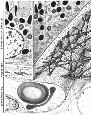
| Fig. 3. Schematic drawing of the inner choroid and retinal pigment epithelium (RPE). The villi (a) of the RPE extend internally to enclose the outer segments (b) of the photoreceptors. The intercellular junctions are characterized by a zonula occludens (c) and a macula adherens (d). The cytoplasm of the RPE contains a nucleus (e), mitochondria (f), a Golgi apparatus (g), melanin granules (h), phagocytosed outer segment tips (i), and smooth endoplasmic reticulum (j). The plasma membrane shows basal infoldings (l) and a basal lamina (m). In addition to the RPE basal lamina, Bruch's membrane comprises inner and outer collagenous layers (o and p), an elastin layer (n), and the basal lamina (m) of the choriocapillaris. The choriocapillaris (q) has a fenestrated epithelium (r, arrows). The intercapillary zone contains collagen (s). The lumen of the capillary contains two erythrocytes. (Hogan MJ, Alvarado JA, Weddell JE: Histology of the Human Eye. Philadelphia: WB Saunders, 1971.) |