|
|
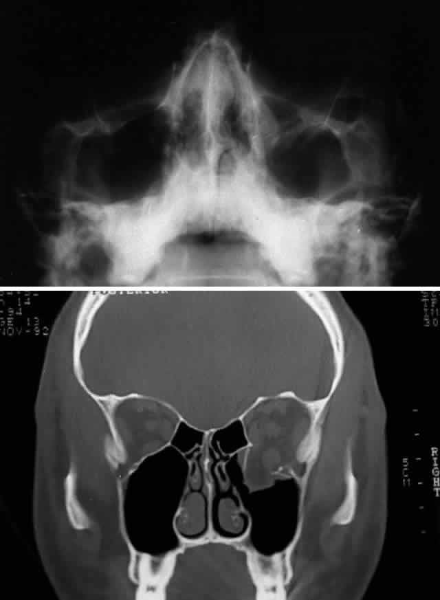
| Fig. 4. Same patient as depicted in Figure 3. A. Water's view. Facial x-ray shows ill-defined left inferior orbital floor fracture with soft tissue mass in superior left maxillary sinus. B. Coronal CT scan shows superior resolution of left orbital floor defect, with prolapse of orbital soft tissues into the maxillary sinus. Note distortion of left inferior rectus muscle. Floor defects greater than 50% are more likely to produce enophthalmos. |