|
|
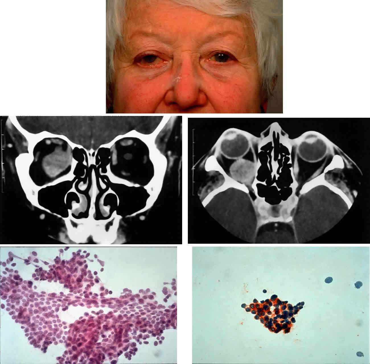
| Fig. 19. Case 1: Mass syndrome of presentation. Clinical appearance: proptosis, upward displacement of the globe, and mechanical restriction of ductions (A). CT scan shows an inhomogeneously enhancing retrobulbar mass (B) displacing the optic nerve superiorly and inseparable from the inferior rectus muscle (C). Needle aspiration biopsy showed cohesive groups of tumor cells (D) (H & E, × 320) that stain positively for chromogranin on immunohistochemistry (E) (H & E, × 320), which is consistent with a metastatic carcinoid tumor. |