|
|
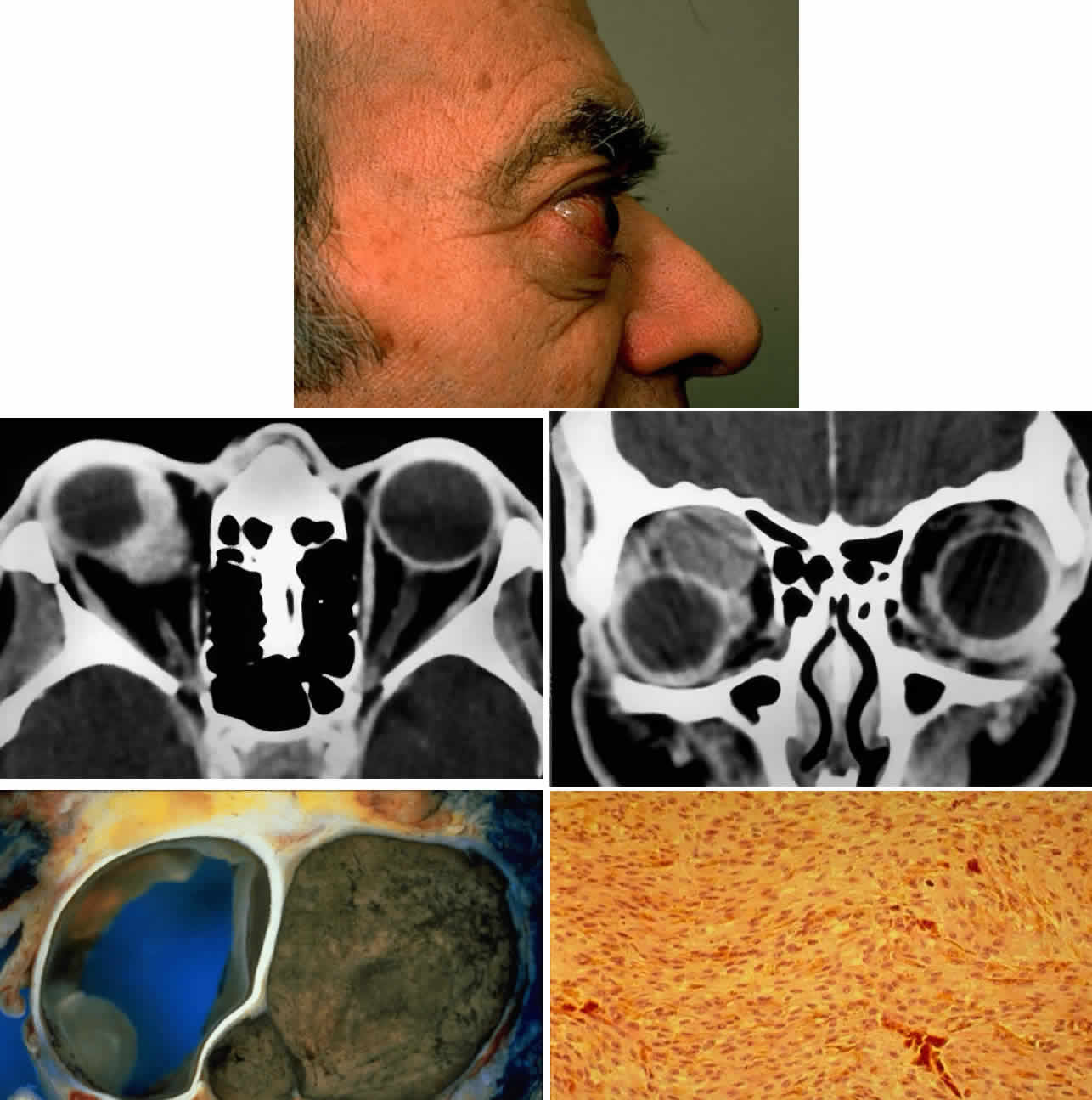
| Fig. 13. A 65-year-old man presented originally with a right retinal detachment and secondary glaucoma due to a large choroidal melanoma. The patient refused enucleation and self-treated instead with herbal medicines and “health foods.” He presented again 6 years later with acute onset of proptosis associated with pain, periocular edema, erythema, and marked restricted ductions (A). CT scan revealed the intraocular tumor, with massive orbital extension and displacement of the globe anteriorly, downward, and laterally (B and C). He underwent lid-splitting exenteration and split-thickness skin graft from the anterior thigh. The gross specimen shows the massive orbital component of melanoma, which had extended from the posterior choroid through emissarial vessels (D). An area of predominantly spindle-shaped melanoma cells with scattered, heavily pigmented macrophages is shown (E) (H & E, × 200). (D and E courtesy of Valerie A. White, MD, Department of Pathology, University of British Columbia, and the Vancouver Hospital and Health Sciences Center.) |