|
|
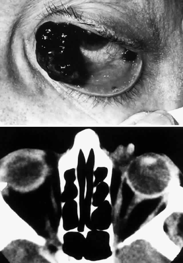
| Fig. 12. An 81-year-old white woman had been aware of a pigmented lesion in the left caruncular region for 18 months. It had gradually increased in size and had bled on several occasions. In addition, she was aware of a lump on the left side of her neck for 6 weeks. On examination, a large, nodular, pigmented medial conjunctival lesion associated with thickening and induration of the tarsal conjunctiva was noted, as was a nodular satellite lesion in the temporal aspect of the inferior fornix due to local lymphatic spread (A). There was lateral displacement of the globe and some limitation of abduction. Axial CT scan demonstrates the irregularly shaped mass in the anterior orbit involving the insertion of the medial rectus muscle (B). The patient refused exenteration and underwent local resection, cryotherapy, mucous membrane graft, and radical neck dissection. Of 31 neck nodes removed, 2 were positive for metastatic disease. Nodular recurrence developed in her left jaw, and the patient died 1 year after presentation without evidence of local ocular recurrence. (Rootman J, Ragaz J, Cline R, Lapointe JS: Metastatic and secondary tumors of the orbit. In Rootman JR (ed): Diseases of the Orbit: A Multidisciplinary Approach, pp 405–427. Philadelphia, JB Lippincott, 1988.) |