|
|
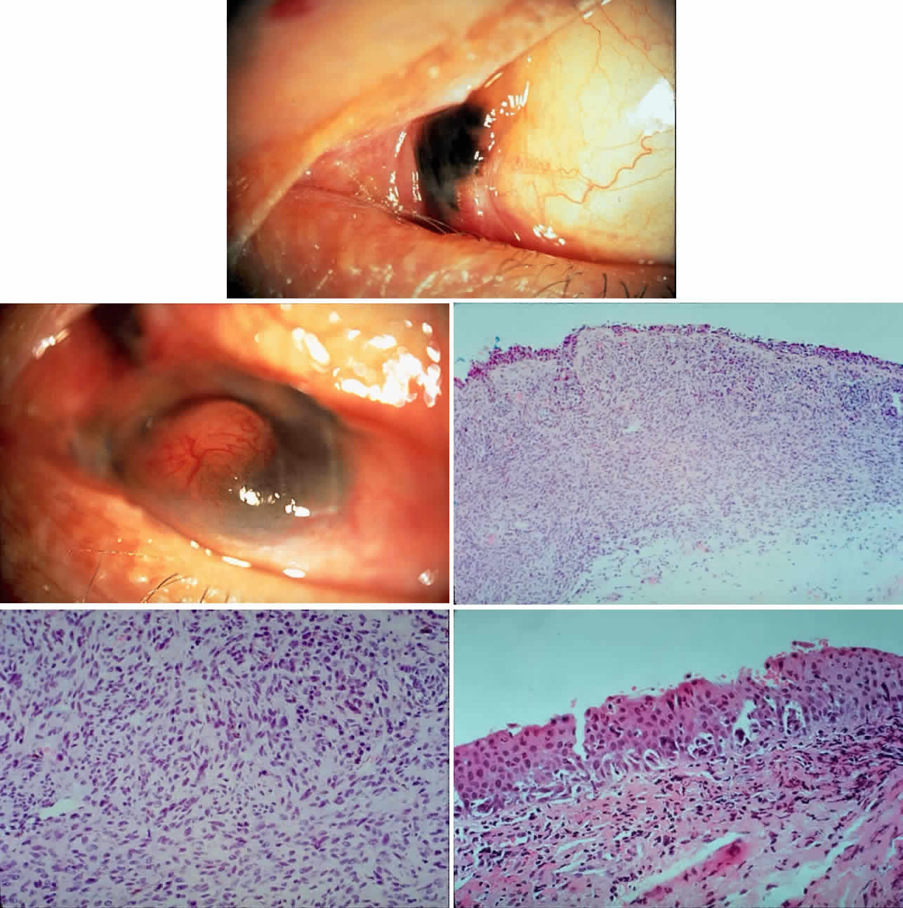
| Fig. 11. A 61-year-old white man noted a pigment spot in the medial canthal area of his left eye associated with intermittent subconjunctival hemorrhages lasting 6 months. On examination, there was a raised, pigmented lesion in the lower medial conjunctiva (A) measuring 20 mm across the base, with a central, pale, telangiectatic nodule 6 mm thick noted on lateral gaze (B). There was extension into the upper medial fornix and laterally in the lower fornix. There was no evidence of preauricular, submandibular, or cervical lymphadenopathy. A clinical diagnosis of conjunctival melanoma arising in primary acquired melanosis (PAM) was made. Conjunctival biopsy revealed invasive melanoma (C and D) (H & E, × 80 and × 200, respectively) arising within PAM type IB (E) (H & E, × 200). A total exenteration and split-thickness skin graft from the anterior thigh were performed, and the patient was disease-free at 6 months' follow-up. |