|
|
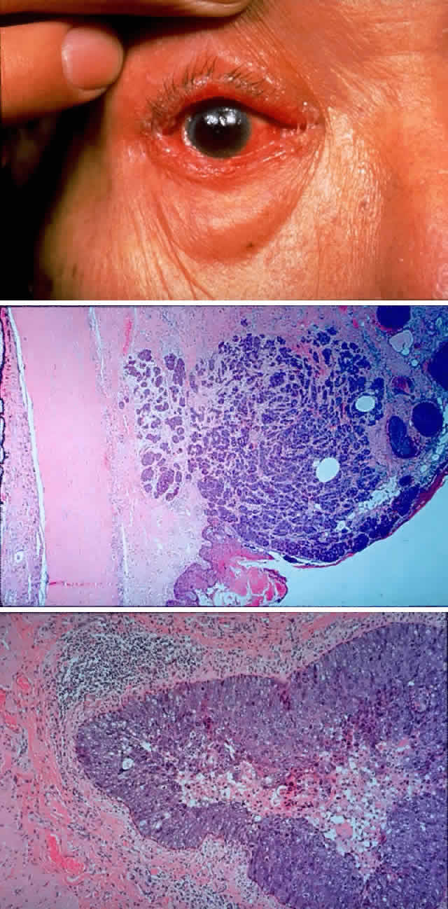
| Fig. 9. A 79-year-old man had been treated for several years for chronic blepharoconjunctivitis. On examination, he had thickening of upper and lower lid margins, diffuse symblepharon, conjunctival thickening, injection, and yellowish plaque-like foci (A). Conjunctival biopsy confirmed invasive sebaceous cell carcinoma, and subtotal exenteration was performed. Tumor invasion along the globe (B) (H & E, × 32) and into muscle (C) (H & E, × 80) was noted. |