|
|
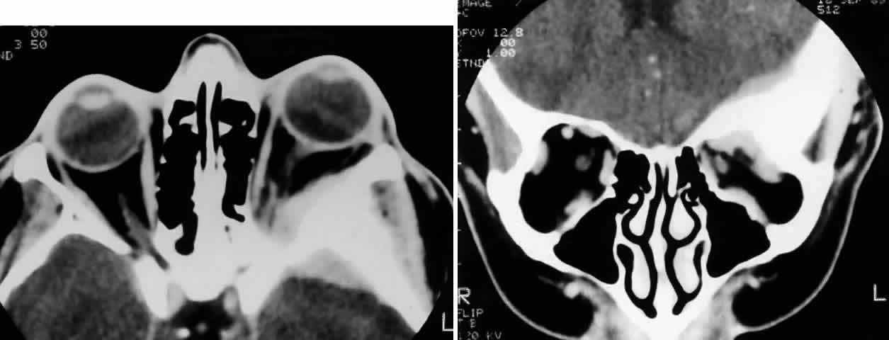
| Fig. 4. A 36-year-old white woman presented with a 3-month history of progressive left proptosis associated with a retrobulbar pressure sensation. On external examination, she had bossing of the temporalis fossa and a proptosis of 9 mm axially, with a slight downward displacement of the globe. Ocular ductions were full, and there was no evidence of optic nerve compromise. CT scan on axial (A) and coronal (B) views confirmed a hyperostosing sphenoid wing meningioma with soft tissue components in the posterolateral orbit and anterior middle cranial fossa. During the next 20 months, her proptosis progressed to 11 mm, accompanied by increasing headaches and ocular pain, ocular restriction, and blurred vision in abduction. She underwent tumor resection with a combined frontotemporal orbitozygomatic approach, and she remains without evidence of recurrence 5 years after surgery. Visual function was preserved, and proptosis decreased to 2 mm. |