|
|
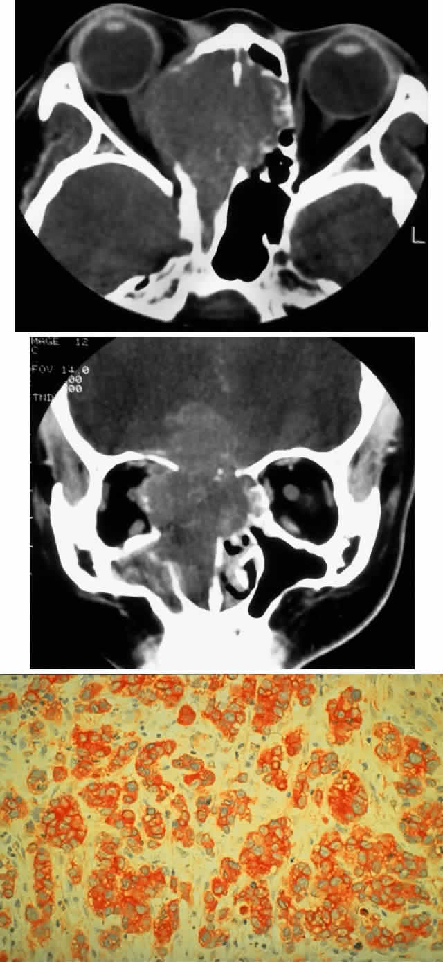
| Fig. 2. A 32-year-old woman presented with a 1-month history of sinus congestion and nasal obstruction unresponsive to antibiotics. In the 1 week preceding the initial exam, the right eye became progressively more prominent and showed tearing and redness. On examination, there was mild edema of the lower lid and dysesthesia in the distribution of cranial nerve V2, and the globe was displaced 6 mm laterally and 5 mm anteriorly. There was mild limitation of abduction, chemosis, and nasal choroidal folds. An axial CT scan revealed a diffuse, destructive soft tissue mass centered in the superior nasal passage and ethmoid sinuses extending into the right orbit in contiguity with the medial rectus (A). On enhanced coronal view, the tumor was noted to involve the right maxillary antrum and to extend superiorly into the anterior cranial fossa (B). Biopsy of the sinus (C) revealed an alveolar-type rhabdomyosarcoma (muscle stain, × 320). |