|
|
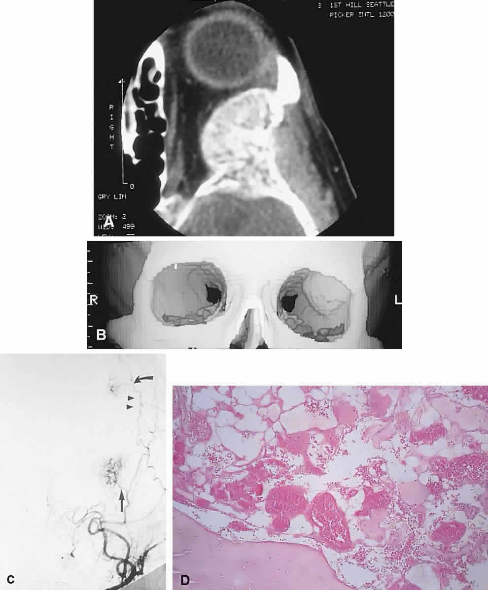
| Fig. 13. A 65-year-old woman with slowly progressive left proptosis. A. CT showed a well-defined, expansile lesion of the greater wing of the sphenoid, which demonstrated a spiculated internal pattern. B. A 3D CT illustrated the expansion of the bone of the lateral orbital wall. C. Subtraction angiography revealed a tangle of vessels at the site of the orbital mass (arrow) in addition to a smaller, more superior lesion in the frontal bone (curved arrow). After preoperative embolization, the mass was excised with a rim of normal bone. D. Histology showed an intraosseous hemangioma with large cavernous, endothelial-lined, blood-filled spaces (hematoxylin-eosin, × 20). (C from Rootman J, Kao SCS, Graeb DA: Multidisciplinary approaches to complicated vascular lesions of the orbit. Ophthalmology 9:1440–1446, 1992.) |