|
|
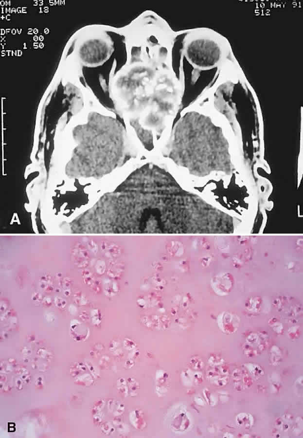
| Fig. 10. This 46-year-old man with a history of midline chondrosarcoma resected 3 years previously presented with a right optic neuropathy. A. CT revealed recurrent tumor in the form of a heterogeneous mass involving both ethmoidal sinuses and orbit. Mottled areas of mineralization are apparent in the matrix of the lesion. B. The tumor was resected through an orbitocranial approach, and the histology revealed a grade 2 chondrosarcoma composed of hypercellular cartilage with lacunae containing binucleated chondrocytes (hematoxylin-eosin, × 50). He remains free of recurrence after 4 years. |