|
|
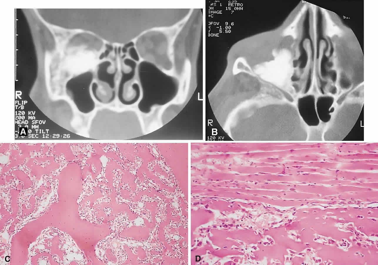
| Fig. 9. A painful right inferior orbital mass effect developed over several weeks in this 19-year-old man with a history of bilateral retinoblastoma treated with radiation therapy and chemotherapy. A and B. CT showed an irregular, largely sclerotic mass involving the orbital floor. He was treated with an en bloc orbitectomy and chemotherapy but died 4 years later from acute myeloid leukemia. C. Histology revealed a sarcomatous stroma with osteoid production forming lacelike patterns (hematoxylin-eosin, × 20). D. A high-power view of the sarcoma adjacent to extraocular muscle indicates that the anaplastic cells appear less atypical with incorporation into the osteoid, the so-called normalization of malignant osteoid (hematoxylin-eosin, × 50). |