|
|
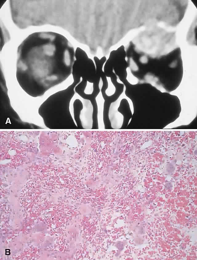
| Fig. 8. A 69-year-old woman with acutely painful left proptosis for 2 days. Examination showed left preseptal edema associated with 9 mm of proptosis and 7 mm of inferior displacement. A. CT showed a well-defined, mottled superior orbital mass with erosion of the roof. B. Histology demonstrated hemorrhage associated with numerous osteoclastic giant cells, in addition to new bone formation (hematoxylin-eosin, × 50). |