|
|
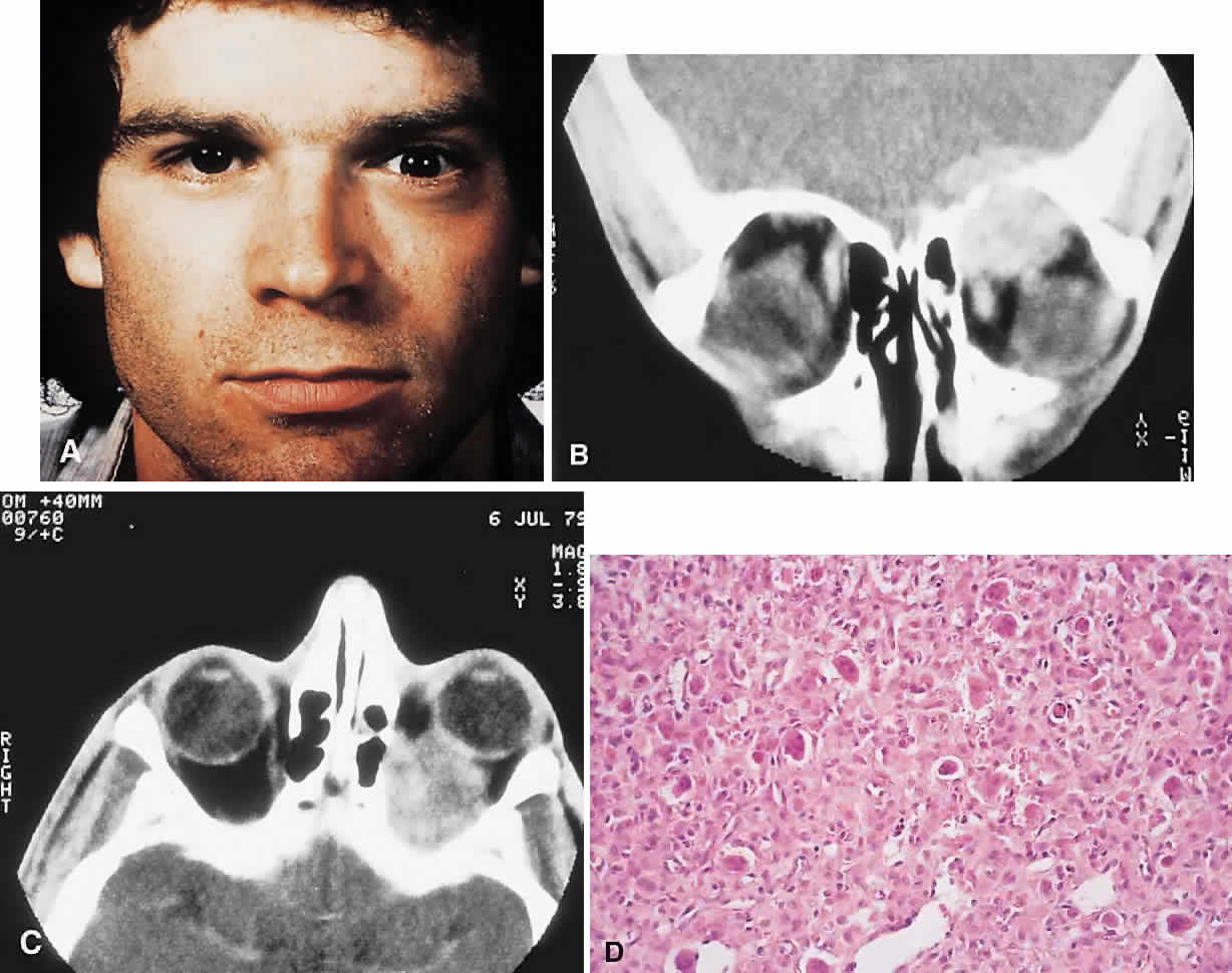
| Fig. 7. A 22-year-old man experienced left proptosis over several weeks. A. On examination, he had 7 mm of proptosis with 3 mm of lateral and inferior globe displacement. B and C. CT revealed a heterogeneous superior orbital mass with destruction of the roof, leading to extradural extension. D. A frontal craniotomy was performed, and histology showed a giant cell granuloma with a fibrous stroma containing plump fibroblasts and giant cells (hematoxylin-eosin, × 50). There was no recurrence at follow-up 4 years later. (B from Rootman J: Diseases of the Orbit: A Multidisciplinary Approach, p 369. Philadelphia: JB Lippincott, 1988.) |