|
|
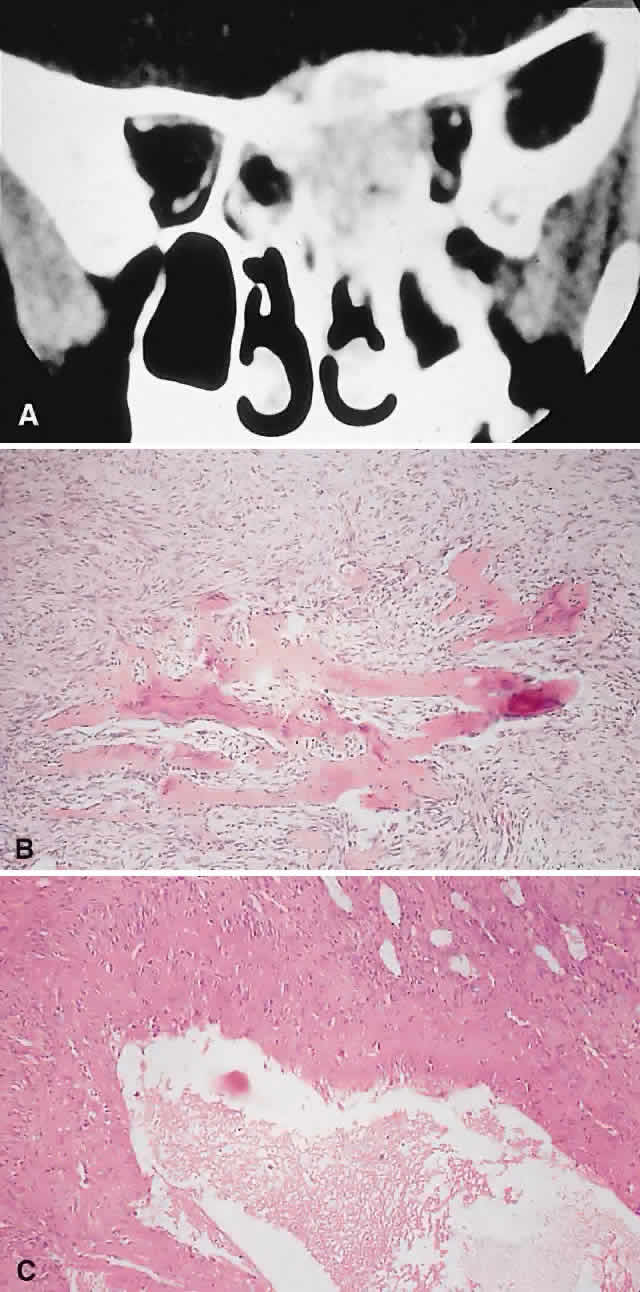
| Fig. 6. An 11-year-old girl with a past history of acute lymphoid leukemia presented with decreasing left vision and proptosis. She had a left best corrected vision of 20/30, a relative afferent pupillary defect, and 2 mm of proptosis. A. CT demonstrated a destructive heterogeneous mass arising in the posterior ethmoid and sphenoid sinuses and involving the left orbital apex. A subtotal removal of the mass was achieved through a frontal craniotomy and orbitotomy. B. Histology showed a fibrous stroma containing giant cells, lymphocytes, and trabeculae of osteoid and bone (hematoxylin-eosin, × 20). There were also areas with giant cells, hemosiderin-laden macrophages, and small foci of aneurysmal sinusoids, leading to a diagnosis of solid aneurysmal bone cyst. There was no evidence of recurrence at follow-up 6 years later. C. Histology from another patient with aneurysmal bone cyst shows a typical cavernous, blood-filled space lacking endothelial lining, pericytes, or smooth muscle (hematoxylin-eosin, × 20). (A from Rootman J: Diseases of the Orbit: A Multidisciplinary Approach, p 373. Philadelphia: JB Lippincott, 1988.) |