|
|
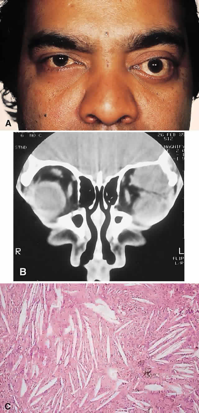
| Fig. 5. A 41-year-old man had a 2-year history of left proptosis and headaches. A. Examination found 6 mm of proptosis with 4 mm of inferior ocular displacement. B. CT showed an osteolytic mass arising from the superolateral frontal bone and extending into the orbit. C. Histologically, a classic picture of cholesterol granuloma was apparent, with numerous cholesterol clefts surrounded by granulomatous inflammation containing foreign body giant cells (hematoxylineosin, × 20). |