|
|
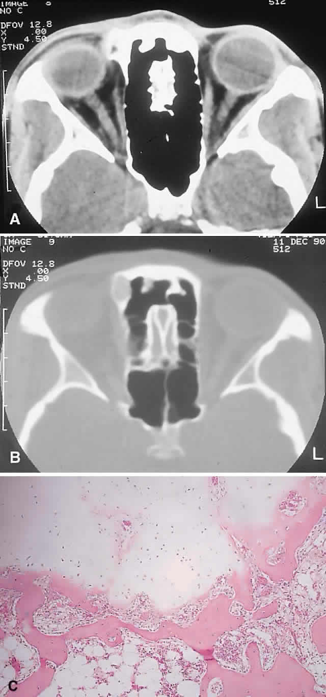
| Fig. 4. This 25-year-old woman had a 6-month history of a hard lump, which was readily palpable at the superomedial right orbital rim. A and B. CT showed a 1-cm bony mass that was more radiolucent centrally on bone window setting. C. The histopathology consisted of globules of mature cartilage encased in lamellar bone, leading to a diagnosis of enchondroma (hematoxylin-eosin, × 20). |