|
|
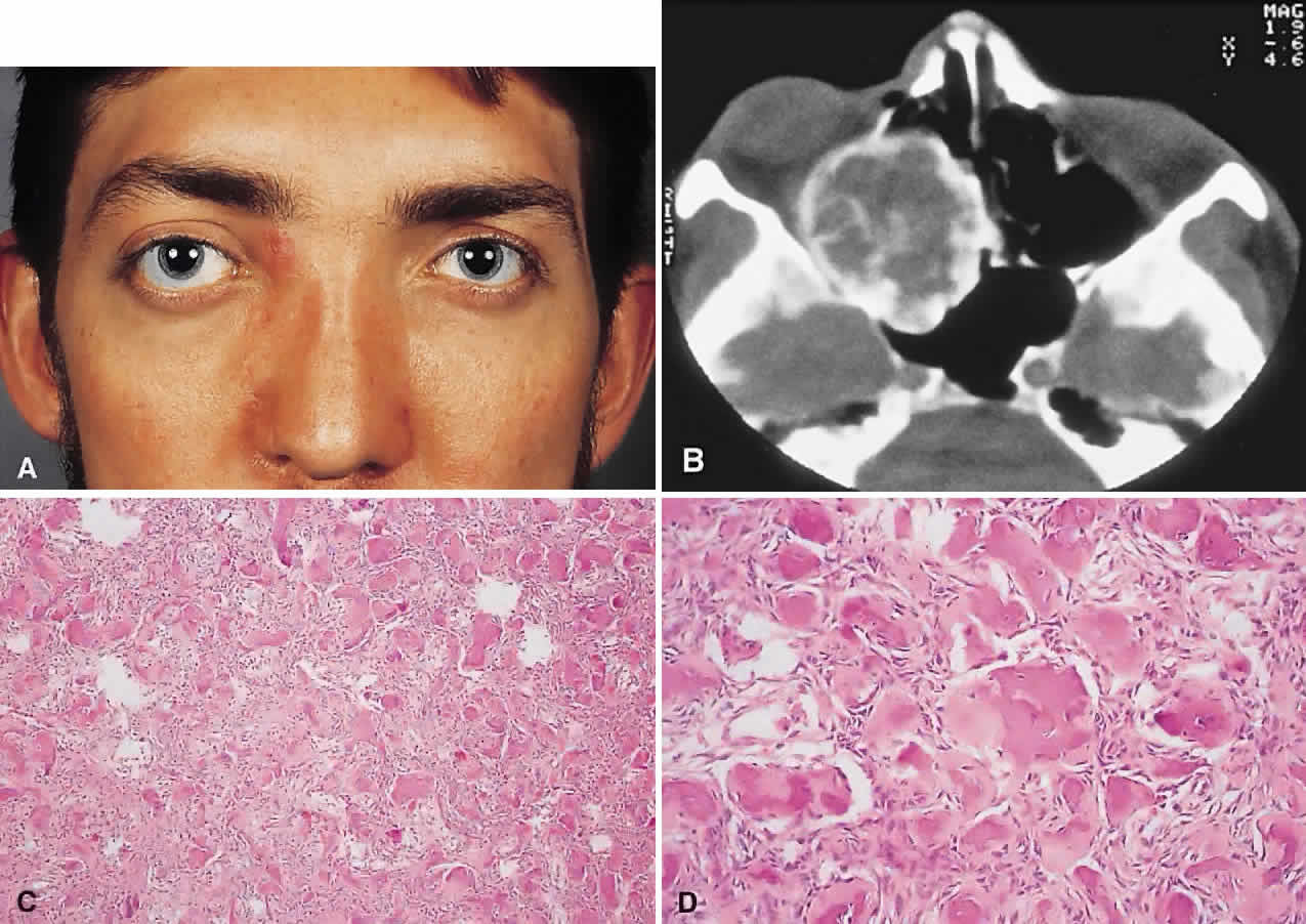
| Fig. 3. A. A 24-year-old man with a 10-year history of increasing right proptosis and a history of ossifying fibroma excised from the right ethmoid and sphenoid 12 years previously. Examination revealed 3 mm of proptosis and 2 mm of lateral globe displacement. B. CT showed a heterogeneous mass with a sclerotic margin involving the right ethmoid and orbit. C and D. After excision, the histology revealed a fibrous stroma containing small spherical ossicles characteristic of the psammomatoid variant of ossifying fibroma (hematoxylin-eosin; C × 20, D × 50). There has been no recurrence in 18 years of follow-up. |