|
|
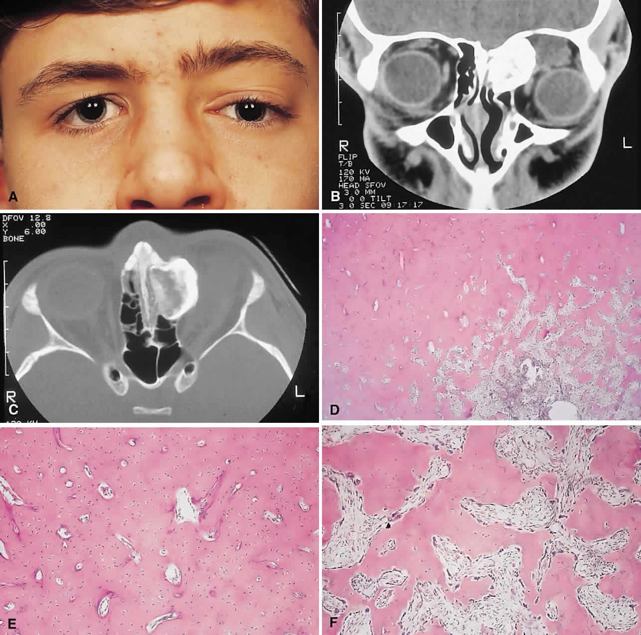
| Fig. 1. A. A 15-year-old boy with a 10-month history of left proptosis had an inferolaterally displaced globe and 6 mm of proptosis. B. CT scan showed a circumscribed sclerotic mass arising from the frontoethmoidal area, with a frontal mucocele laterally. C. Bone windows revealed the bony mass to have a cancellous core and a sclerotic periphery. D. The histology showed a peripheral zone of compact bone, with increasing osteoblastic activity and fibrous tissue toward the center (hematoxylin-eosin, × 5). E and F. Higher power revealed areas of compact bone with haversian canals similar to normal cortical bone and regions of trabecular bone (F) with more osteoblastic activity present (hematoxylin-eosin, × 20). |