|
|
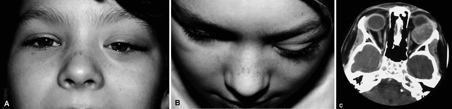
| Fig. 1. A. Clinical photograph depicting proptosis of the left eye with inward and downward displacement and fullness of the superior sulcus. Note absence of inflammatory signs. B. Clinical photograph, superior view, of the same patient. C. Axial computed tomography of the same patient demonstrating a well-defined mass in the superior temporal orbit. A biopsy confirmed embryonal rhabdomyosarcoma. (Courtesy of James Garrity, MD, Mayo Clinic, Rochester, Minnesota.) |