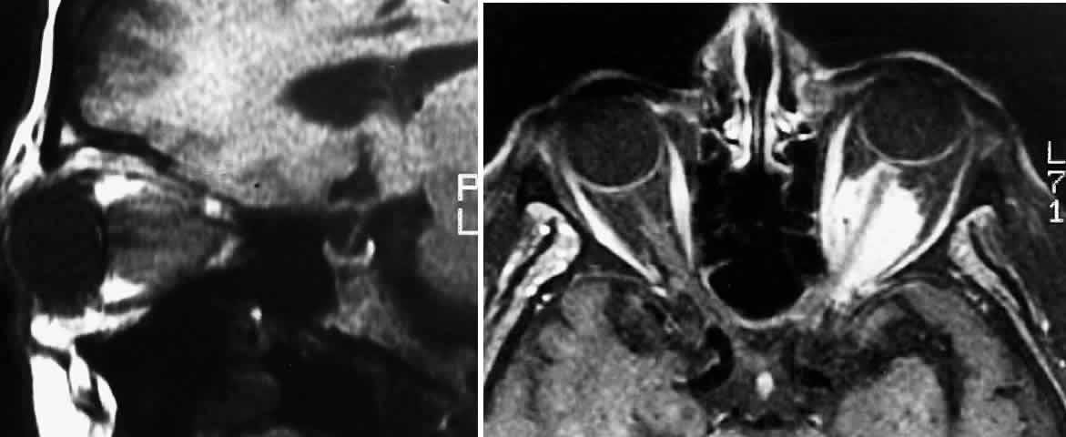
Fig. 5.
A.
T1-weighted oblique MRI of the orbit, demonstrating an optic nerve glioma.
B.
Contrast-enhanced T1-weighted axial MRI of the orbit, demonstrating left meningioma with intracanalicular extension.