|
|
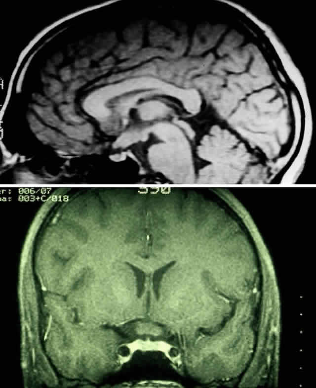
| Fig. 4. A. T1-weighted sagittal MRI of a patient with neurofibromatosis type 1, demonstrating enlarged optic chiasm consistent with optic glioma. B. T1-weighted coronal image with gadolinium and fat suppression in the same patient, demonstrating the chiasmal glioma. |