Fig. 2.
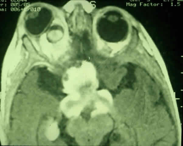
Contrast-enhanced T1-weighted axial MRI of the orbits of the patient in
Figure 1
, demonstrating a large optic pathway glioma with posterior extension into the optic tracts and radiations. (Courtesy of Orlando Ortiz, MD, and Jeffrey Hogg, MD)