|
|
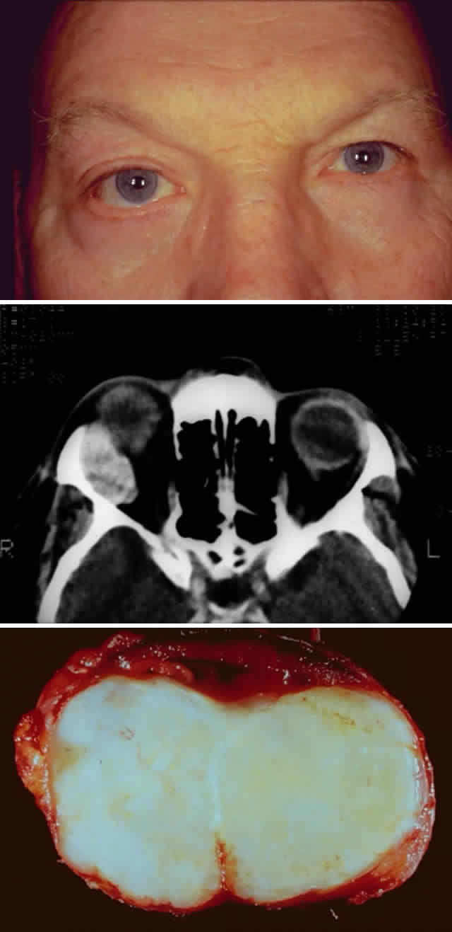
| Fig. 4. A 47-year-old man with a 2-year history of slowly progressive, nonaxial proptosis secondary to a benign pleomorphic adenoma. A. Clinical photograph demonstrating proptosis of the right eye with asymmetric fissures and mild inferior and medial displacement of the globe. B. Computed tomographic image of the pleomorphic adenoma. Note the smooth bone erosion of the lacrimal fossa. The lesion does not extend beyond the orbital rim. C. Photograph of the gross specimen, which has been cut to demonstrate its solid consistency contained within a “pseudocapsule.” |