|
|
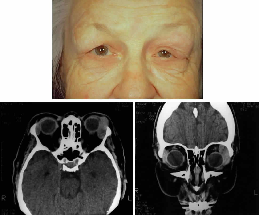
| Fig. 2. A 68-year-old woman with malignant lymphoma. A. Clinical photograph demonstrating fullness over the left lacrimal gland fossa, S-shaped contour of the upper eyelid, asymmetric superior sulci, and ptosis. Inflammatory signs are absent. B. Axial computed tomograph demonstrating the classic well-demarcated, oblong appearance of lymphoproliferative diseases of the lacrimal gland. Note that the lesion extends beyond the anterior orbital rim. C. Coronal computed tomograph of the same lesion. Note that the lesion contours to the globe and bone and does not produce any bone changes. |