|
|
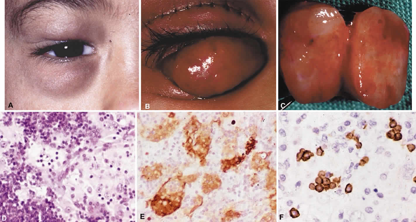
| Fig. 33. A. A child with sinus histiocytosis with massive lymphadenopathy involving the right upper and lower preseptal region. B. Intraoperative photo showing the location of the encapsulated well-circumscribed lesion. C. Surgical specimen incised to demonstrate the yellow-red variegated appearance. D. Histologic study showing a small lymphoid infiltrate between sheets of large, pale-staining histiocytes with round vesicular nuclei and prominent nucleoli. Some of these histiocytes appear to contain small lymphocytes within their cytoplasm (hematoxylin and eosin, × 10). E. CD68 staining confirms the presence of large histiocytes. Within their cytoplasm they contain numerous nonstaining lymphoid cells which are undergoing lymphophagocytosis or emperipolesis (immunoperoxidase, × 40). F. CD20 staining confirms the presence of clusters of small B cells within the cytoplasm of the pale histiocytes (immunoperoxidase, × 60). |